FIG. 7.
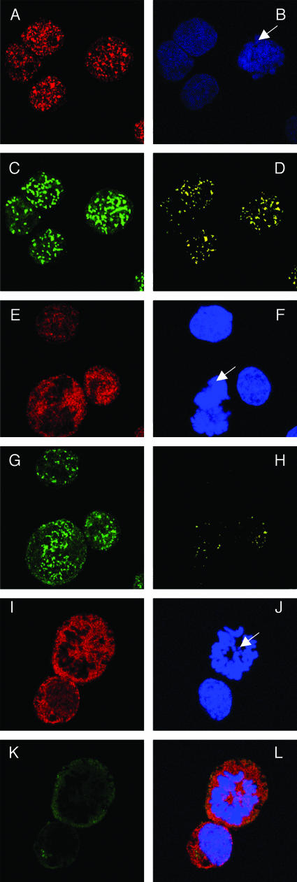
Colocalization of Brd4 and LANA on mitotic chromosomes in KSHV-positive BCLM cells. (A to D) BCLM cells were double stained with N-MCAP (A) and LN53 (C) as described in the legend to Fig. 6. Cells were also counterstained with DAPI (B) (the arrow indicates a mitotic chromosome). Colocalization of Brd4 and LANA was distinctly observed on mitotic chromosomes as bright yellow dots (D). (E to H) BCLM cells were double stained with an anti-Brd2 peptide rabbit polyclonal antibody and LN53. The staining was detected by incubation with a Alexa Fluor 594 goat anti-rabbit IgG (E) and a goat anti-rat IgG FITC conjugate (G), respectively. The staining of DAPI is shown in panel F. The overlay signal of panels E and G is shown in panel H. In contrast to the LANA staining, a majority of Brd2 staining is excluded from the mitotic chromosomes (E and H). (I to L) KSHV-negative BJAB cells were double stained for Brd2 and LANA as for panels E to H. The overlay signal is shown in panel L. In contrast to the status in KSHV-positive cells, the Brd2 staining is almost completely excluded from mitotic chromosomes.

