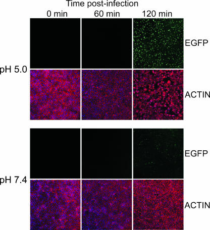
FIG. 2.
Synchronization of infection by low-pH treatment. BS-C-1 cell monolayers were passively inoculated with EGFP-expressing MVs at a multiplicity of 5 PFU per cell at 4°C for 1 h. After being washed, cells were treated with pH 5.0 or 7.4 buffer for 3 min at 37°C, washed and incubated in EMEM for 0, 60, or 120 min at 37°C, fixed with 3% paraformaldehyde, and quenched with 2% glycine. Cells were stained with Alexa Fluor 568-conjugated phalloidin to visualize filamentous actin and examined by confocal microscopy. Actin, red; EGFP fluorescence, green.

