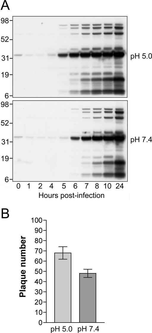
FIG. 3.
Low-pH treatment enhances a productive virus infection. (A) Western blots comparing VACV late gene expression following low- and neutral-pH treatments. BS-C-1 cell monolayers were incubated with purified MVs at 5 PFU per cell at 4°C for 1 h. After being washed, cells were treated with pH 5.0 or pH 7.4 buffer for 3 min at 37°C, washed with EMEM, and incubated at 37°C. At the indicated times, cell lysates were analyzed by sodium dodecyl sulfate-polyacrylamide gel electrophoresis, transferred to nitrocellulose, and analyzed by Western blotting with an anti-VACV antiserum. The positions and molecular masses in kDa of marker proteins are shown on the left. (B) Effect of pH treatment on plaque formation. BS-C-1 cell monolayers in six-well plates were inoculated with approximately 50 PFU per well of purified MVs at 4°C for 1 h. After removal of unbound virus by washing, cells were treated with pH 5.0 or pH 7.4 buffer for 3 min, washed with EMEM, overlaid with 2% methylcellulose in EMEM, and incubated at 37°C. At 48 h postinfection, monolayers were fixed and stained with crystal violet and plaques counted. Experiments were performed in triplicate, and data points represent the means ± standard errors.

