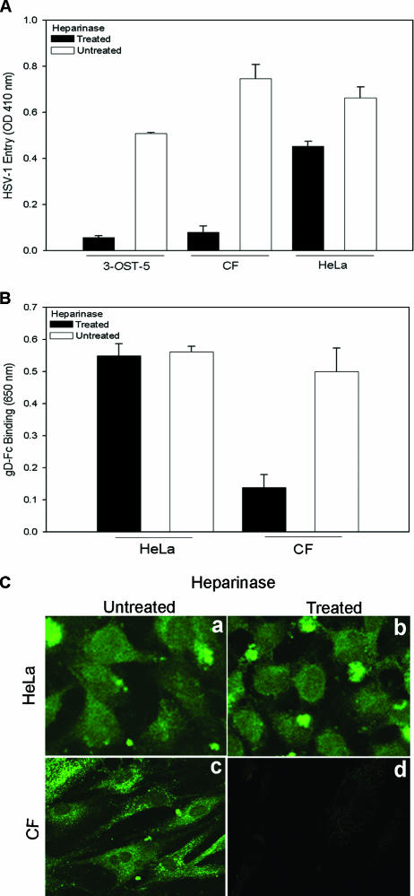
FIG. 4.
Effect of heparinase treatment on HSV-1 entry and gD binding to CF. (A) Effect of heparinase treatment on HSV-1 entry. Cultured CF, HeLa cells, or control CHO-K1 cells expressing 3-OST-5 were treated with heparinases II and III (4 U/ml) in 96-well tissue culture dishes. A separate set of cultured cells were mock treated with PBS alone. The cells were then exposed to HSV-1(KOS) gL86 at 35 PFU/cell, and viral entry was quantitated 6 h later, as described in the legend to Fig. 1A. Data represent the mean ± the standard deviation of results in triplicate wells in a representative experiment. The experiment was repeated three times with similar results. (B) Effect of heparinase treatment on gD binding. Cultured CF and control HeLa cells in 96-well dishes were treated with heparinases II and III (4 U/ml) or incubated with buffer. The cells were then incubated with gD-Fc for 30 min, followed by fixation and incubation with a secondary antibody and a horseradish peroxidase detection system. The values shown (means of triplicate determinations plus standard deviations) represent the amounts of binding product detected spectrophotometrically (OD650). (C) Confocal imaging of gD binding to heparinase-treated cells. Cultured HeLa cells and CF were plated in glass bottom culture dishes and grown overnight. The cells were treated with heparinases II and III (4 U/ml) or mock treated with buffer. After incubation with gD-Fc, the cells were washed, fixed with acetone, and incubated with FITC-conjugated anti-rabbit IgG. Micrographs of representative HeLa cells and CF are presented to show gD-Fc binding with or without heparinase treatment with a Leica confocal microscope.

