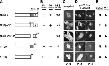
FIG. 3.
Vp3 D1 directs cytoplasmic Vp1 interaction. TC-7 cells grown on coverslips were injected with the respective pSGVp3 constructs with or without pSGVp1. (A) Schematic diagrams of pSGVp3 constructs in which regions are deleted from the mutant Vp3s are shown. X indicates mutations of Vp3 NLS 201-KKKRK-205 to NNNGN. (B) Locations of Vp3 mutations and deletions. mut, mutation; dl, deletion. Other abbreviations are defined in the legend to Fig. 1B. (C) Subcellular localization of mutant Vp3s following microinjection of the respective pSGVp3 constructs. (D) Each set of photographs shows subcellular localization of Vp3 on the left and Vp1 on the right, in the cells injected with a mixture of the respective pSGVp3 and pSGVp1 mutants. (E) Summary of the subcellular localization of Vp3 mutants and coexpressed Vp1. N, nuclear localization; C, cytoplasmic localization.

