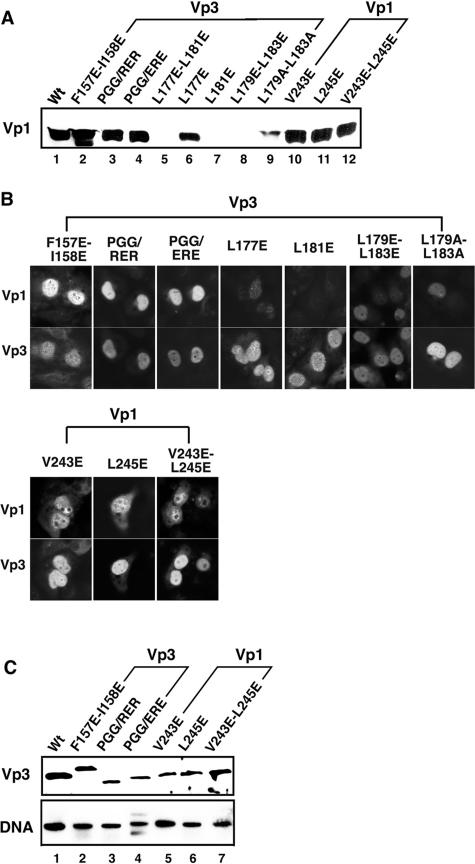
FIG. 6.
Levels of capsid proteins and viral DNA in cells transfected with mutant DNAs. (A) Level of Vp1. Lysates of the cells transfected with either wild-type or mutant NO-SV40 DNA were examined for Vp1 by immunoblotting. Approximately 1/200 of the lysates of cells transfected with wild-type DNA (lane 1), Vp3 mutants (lane 2, F157E-158E; lane 3, PGG/RER; lane 4, PGG/ERE; lane 5, L177E-L181E; lane 6, L177E; lane 7, L181E; lane 8, L179E-L183E; lane 9, L179A/L183A), and Vp1 mutants (lane 10, V243E; lane 11, L245E; lane 12, V243E-L245E) were used for immunoblotting. (B) Subcellular localization of capsid proteins. TC-7 cells grown on coverslips were transfected with each viral DNA, fixed at 24 h posttransfection, and then incubated with guinea pig anti-Vp1 and rabbit anti-Vp3 sera, followed by fluorescein- and rhodamine-conjugated secondary antibodies (22). In each pair of photos, the top and bottom panels show identical cells stained for Vp1 (top) and Vp3 (bottom). A set of photographs for cells transfected with Vp3 mutants is bracketed as Vp3, and those with Vp1 mutants are bracketed as Vp1. The cells transfected with Vp3 L177E-L181E did not show positive signs for the capsid proteins and thus are not shown. (C) Levels of Vp3 and viral DNA. The mutants showing normal levels of Vp1 in panel A were tested for level of Vp3 by Western blotting and DNA by Southern blotting. Vp3 mutants F157E-I158E (lane 2), P164R-G165E-G166R (PGG/RER) (lane 3), and P164E-G165R-G166E (PGG/ERE) (lane 4) and Vp1 mutants V243E (lane 5), L245E (lane 6), and V243E-L245E (lane 7) are marked.

