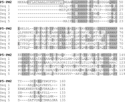
FIG. 6.
Sequence alignment of the structural protein P5 of phage PM2 with similar sequences present in GenBank. Amino acid residues of P5 that are conserved in at least one other protein are shaded gray. A predicted transmembrane helix in the PM2 protein is boxed. P5-PM2, structural protein P5 of bacteriophage PM2 (accession no. NP_049909); Seq 1, Gp21 of bacteriophage D3112 (accession no. NP_938228); Seq 2, putative structural protein P5 of Methylococcus capsulatus prophage MuMc02 (accession no. AAU90959); Seq 3, Orf36 of Photorhabdus luminescens (accession no. AAO18061); Seq 4, conserved hypothetical phage protein of Bacteroides fragilis NCTC 9343 (accession no. CAH07979); Seq 5, conserved hypothetical protein of Desulfovibrio vulgaris subsp. vulgaris strain Hildenborough (accession no. AAS95986). The alignment was constructed using CLUSTALW (11).

