
FIG. 2.
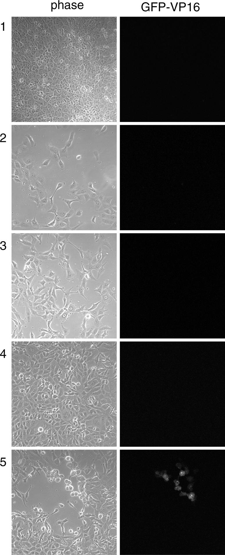
Propagation of an MDBK monolayer in a repressed state of infection. MDBK cells infected for 12 days with 2,000 PFU of HSV-1 [v44] were trypsinized, split (1:20 ratio), and replated in six-well cluster dishes. Cell progression was monitored thereafter by confocal microscopy. Images were taken at 12 dpi (panels 1) and 1 (panels 2), 2 (panels 3), and 3 (panels 4 and 5) days posttrypsinization.