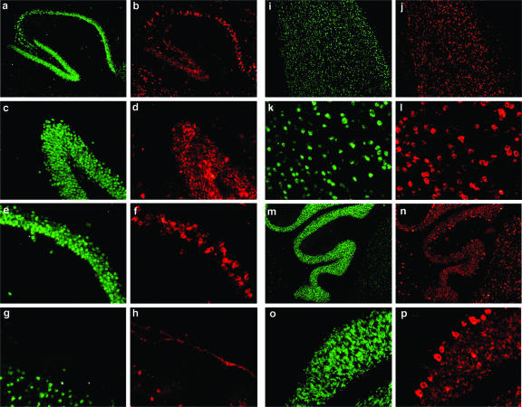
FIG. 2.
Virus distribution in the brain of LCMV-cgPi mice. Viral antigen in whole-brain sections of 6-month-old LCMV-cgPi mice was detected by immunofluorescence staining performed using frozen brain sections and hyperimmune guinea pig serum to LCMV and a rhodamine red X-labeled secondary antibody (red). Neurons were labeled with an antibody to NeuN and a FITC-labeled secondary antibody (green). a and b, hippocampus; c and d, dentate gyrus; e and f, C1 region; g and h, meninges; i to l, cerebral cortex; m to p, cerebellum.

