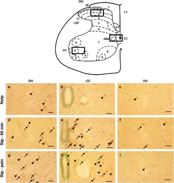
Figure 1.

Immunohistochemistry of FOS+ neurons in the DH, CC, and VH (boxed areas on the schematic of spinal cord) of the L4 spinal cord in nonstepped (first row, a–c), stepped for 60 min (second row, d–f), and pain-associated stepping for 35 min (third row, g–i), rats. FOS+ cells found in the DH, CC, and VH. Scale bar = 10 μm. Arrows = high-intensity staining, arrowheads = low-intensity staining
