
Figure 5.
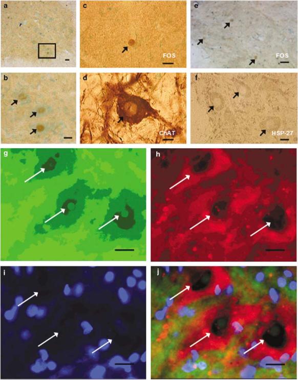
FOS+ motoneurons. Adjacent sections from L4 spinal cord of a Stp rat (60 min) labeled with c-fos, c-fos, and ChAT or HSP-27 and counterstained with methyl green for lamina IX. Distribution of FOS-stained motoneurons in the VH: brown (FOS nuclear stain with DAB), green (cytoplasmic counterstain with methyl green). Scale bar = 40 μm (a). Higher magnification of the same region from (a) with three distinct FOS+ neurons (arrows, b). Scale bar = 20 μm. FOS-stained adjacent section to (d), depicting a clearly labeled nuclear stain of neurons (arrow, c). ChAT section depicting a clear cytoplasmic stain of motoneurons (arrow, d). FOS section stained adjacent to (f) depicting a nuclear stain in three clearly labeled motoneurons (arrows, e). HSP-27 section depicting three motoneurons with clear cytoplasmic staining (arrows, f). Scale bar = 10 μm. Double IF shows FOS+ motoneurons in lamina IX (g–j). FITC – FOS (g); Rhodamine – HSP-27 (h); Dapi – nuclear stain (i); Composite – colocalization of c-fos and HSP-27 (j). Scale bar = 10 μm