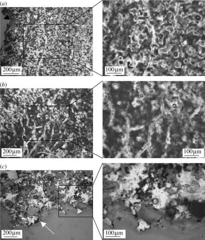
Figure 6.
Light microscopy photomicrographs of ground thin sections of sinter surrounding bird. Insets represent high magnification images of the boxed regions. (a) Ground section of region of sinter immediately adjacent to bird carcass shows evidence of organic substrate, including fungal filaments. Black arrow represents cavity where body of bird was situated. (b) Branching pattern of filaments, consistent with fungi. (c) Ground thin section of region distal to bird body shows obvious decrease in density of sinter fabric and corresponding decrease of organic incorporation. No fungal influence can be seen in this region. White arrow indicates external surface of sinter block.

