
Figure 2.
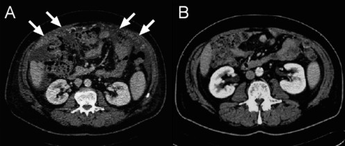
Radiological follow up of peritoneal carcinomatosis. MS- CT axial (contrast-enhanced, portal-venous phase): Peritoneal carcinomatosis (omental cake) (A), regressive under chemotherapy (B).
Official websites use .gov
A
.gov website belongs to an official
government organization in the United States.
Secure .gov websites use HTTPS
A lock (
) or https:// means you've safely
connected to the .gov website. Share sensitive
information only on official, secure websites.
Radiological follow up of peritoneal carcinomatosis. MS- CT axial (contrast-enhanced, portal-venous phase): Peritoneal carcinomatosis (omental cake) (A), regressive under chemotherapy (B).