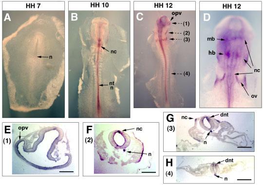
Fig. 2.

Expression of Sema3D in early chick embryos (HH7 to HH12). Dorsal sides of embryos are shown for whole mount in situ hybridization results. For sections, the dorsal sides of the embryons are oriented upward. Whole mount in situ hybridization results are shown for embryos of (A) HH7, (B) HH10, (C) HH12 at a low magnification, and (D) HH12 at a higher magnification. After whole mount in situ hybridization, the embryos were sectioned. The planes of sections were shown with broken arrows and the numbers in parentheses next to the arrows indicate the figures showing the section results. E–H: Results from four section planes (1–4) of the HH12 embryo are shown after in situ hybridization with the Sema3D probe. n, notochord; nc, neural crest; nt, neural tube; mb, midbrain; hb, hindbrain; ov, otic vescicle; opv, optic vescicle; dnt, dorsal neural tube. Scale bars = 100 μm.
