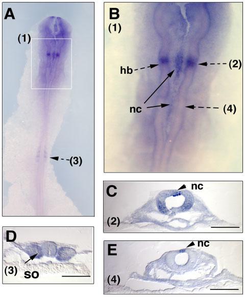
Fig. 3.

Expression of Sema7A transcripts in early chick embryos at HH12. HH12 chick embryos were hybridized with the Sema7A-2 RNA probe, shown at the dorsal side of the embryo (A) and in the hindbrain region (B). The boxed area (1) in A is shown at a higher magnification in B. Note its expression in a restricted domain in hindbrain region and in some premigratory neural crest cells at the dorsal midline of the neural tube. The embryos were sectioned after whole mount in situ hybridization at the indicated section planes (2)–(4). nc, neural crest, hb, hindbrain, so, somite. Scale bars = 100 μm.
