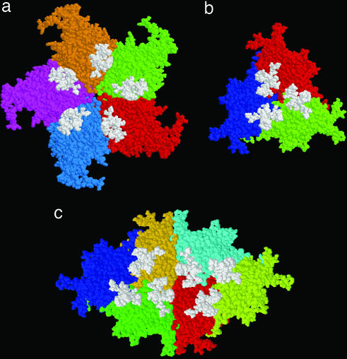
Fig. 3.
Space-filling representations of several symmetry-related MVM capsid subunits (in different colors) and crystallographically observed DNA stretches bound to those subunits (white). Views are from the particle interior and were obtained by using RasMol software (23). (a) Five protein subunits related by a fivefold symmetry axis. (b) Three subunits related by a threefold symmetry axis. (c) Six subunits related by a twofold symmetry axis. Distance measurements show that the center of gravity of each visible DNA stretch is located closer to the capsid twofold axis and further from the fivefold axis.

