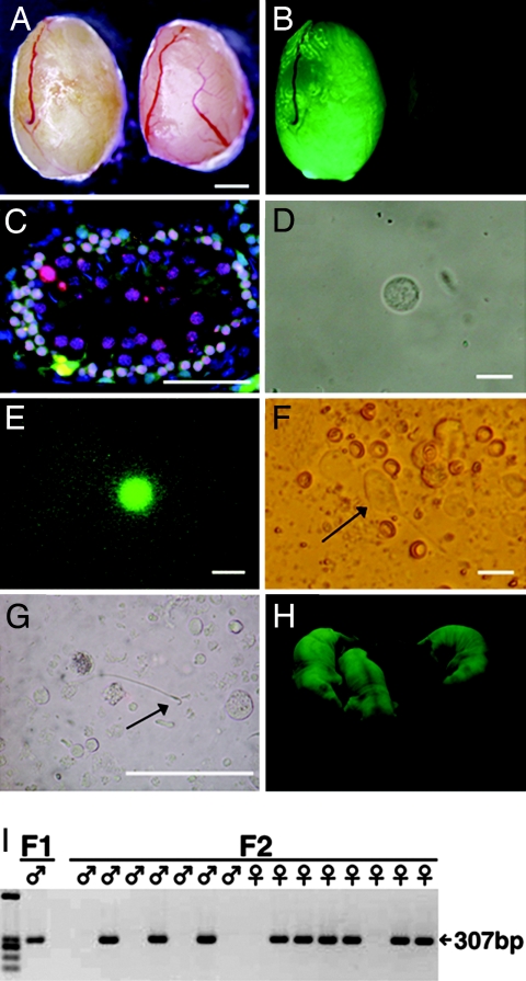
Fig. 1.
Rat spermatogenesis in mouse testis. (A and B) Macroscopic appearance of a nude mouse recipient testis that received EGFP-expressing rat testis cell transplantation (left). The recipient mouse was killed 5 months after transplantation. (B) Note the extensive colonization of donor cells under UV light. Control testis without transplantation did not show fluorescence (right). (C) Histological appearance of a recipient testis that was immunostained with anti-SCP3 antibody (red). The section was counterstained with Hoechst 33258 dye (blue) for nuclei. (D–G) A round spermatid (D and E), elongated spermatid (F, arrow), and spermatozoan (G, arrow) released from a segment of seminiferous tubule that was used to inject eggs. (E) The round spermatid fluoresced under UV light. (H) Rat offspring from a mouse recipient that received cryopreserved rat testis cell transplantation. EGFP expression was observed in the offspring. (I) PCR analysis using DNA extracted from individual F1 and F2 offspring. All EGFP-positive offspring contained the EGFP transgene. (Scale bars: A and B, 1 mm; C, 50 μm; D–F, 15 μm; G, 150 μm.)

