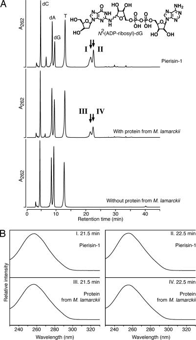
Fig. 2.
Analysis of reaction products formed by DNA, clam protein, and β-NAD. (A) HPLC elution patterns of hydrolysate of DNA incubated with pierisin-1 (Top), with partially purified protein from M. lamarckii (Middle), or without protein (Bottom). These DNA samples were enzymatically hydrolyzed to deoxyribonucleosides and injected into a Develosil RPAQUEOUS column, and the eluate was monitored by measuring its UV absorbance at 262 nm. Arrows indicate the peaks coincident with N2-(ADP-ribos-1-yl)-2′-deoxyguanosine. (B) UV absorption spectra with a photodiode array detector of the compounds (I–IV) in the peak fractions at retention times of 21.5 and 22.5 min presented in A.

