
Figure 2.
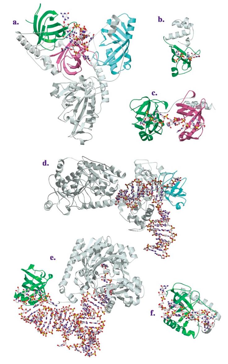
(Continued) Structures of OB-fold/nucleic acid complexes. The high-resolution structures of several OB-fold proteins bound to nucleic acids. The individual OB-fold domains are highlighted in rainbow colors to illustrate the modularity of the domain. (a) OnTEBP ternary complex, (b) EcRho, (c) human RPA, (d) RecG, (e) EcAspRS, (f) Cdc13, (g) EcSSB, (h) L2 in the large subunit of the ribosome, (i) S12 (green), S17 (blue), and IF1 (magenta) in the ribosomal small subunit.