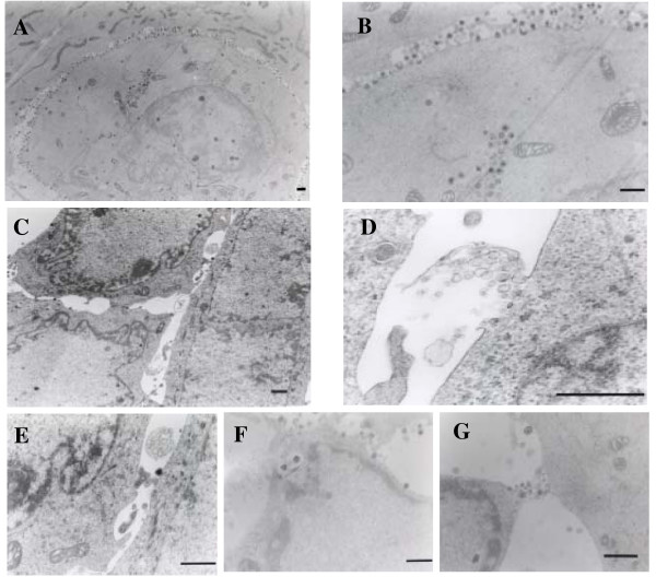
Figure 7.
Electron microscopic observation. FI cells were infected with HSV-1 at an MOI of 2 PFU/cell and cultured for 20 h (A, B). The infected cells were also treated with 1 mM H2O2 for 18 to 20 h p.i. (C to G). To examine cell-to-cell interaction, cultures were fixed in situ and embedded in epoxy resin. Sections were cut parallel to the surface of the dishes. Bar, 1 μm

