
Figure 3.
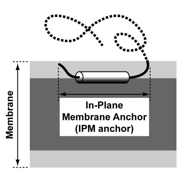
Schematic representation to scale of an IPM anchor. The amphipathic α-helix of the IPM anchor is depicted as a black and white cylinder, for the hydrophobic and hydrophilic sides, respectively. The non-membrane part of the protein is represented by a dotted line. The membrane hydrophobic core, including acyl chains, is dark grey and the membrane interface, including glycerol and above atoms, is light grey.