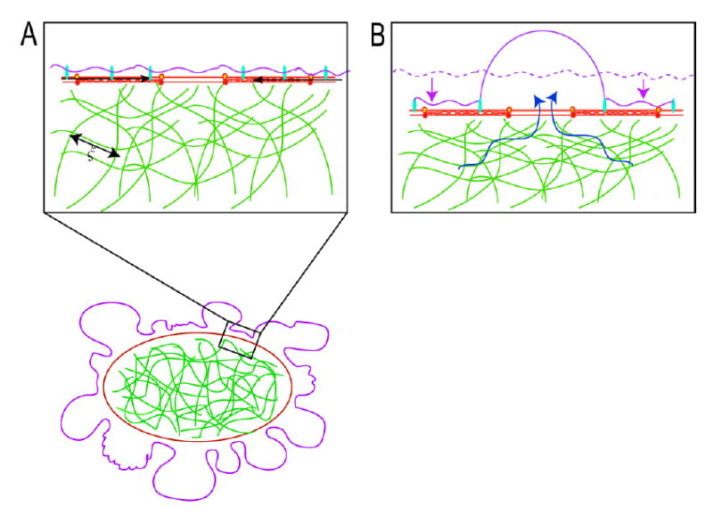
Poroelastic description of blebbing. In all drawings, the actin cortex is drawn in red, the membrane is drawn in mauve, and the cytoskeletal meshwork is drawn in green.
In blebbing cells, a local contraction of the myosin II (black arrows) associated with the actin cortex leads to a shortening of the cortical periphery and therefore to a compression of the cytoskeletal network that fills the cell. The cytoskeletal network is porous and has an average pore size ξ. The compression of the cytoskeletal meshwork creates a hydrostatic pressure in the vicinity of the region of contraction and can lead to bleb nucleation and expansion.
Contraction of the actin cortex leads to a compression of the cytoskeletal network (the dashed mauve line indicates the original position of the cell surface) drives flow of cytosol in the opposite direction (blue arrows). If there is a local defect in membrane-cytoskeleton attachment, a bleb is extruded. Bleb expansion is opposed by two forces: extracellular osmotic pressure and membrane tension.

