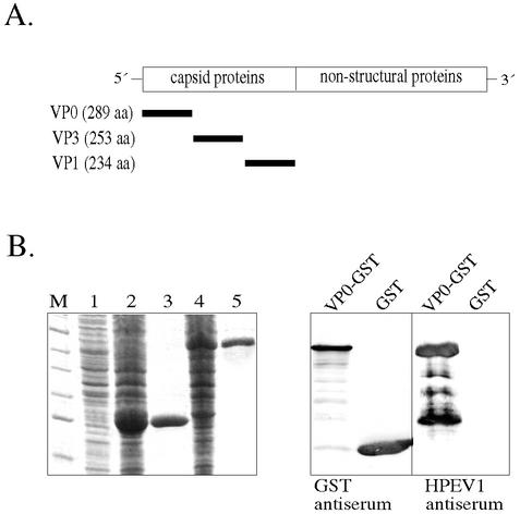
FIG. 1.
(A) Genome localization of the genes for the HPEV1 capsid proteins (VP0, VP3, and VP1) expressed as GST fusion polypeptides. (B) SDS-PAGE (left) and immunoblotting (right) analysis of the VP0-GST fusion protein. Lanes: M, molecular size markers; 1, uninduced E. coli cells; 2, induced E. coli cells expressing GST; 3, purified GST (26 kDa); 4, induced E. coli cells expressing VP0-GST; 5, purified VP0-GST (58 kDa). After separation by SDS-PAGE, the proteins were blotted onto a nitrocellulose filter and detected by primary antibodies (GST antiserum diluted 1:3,000 and HPEV1 antiserum diluted 1:1,000) and secondary antibody (alkaline phosphatase-conjugated anti-rabbit IgG). The samples analyzed in the right panel are VP0-GST and GST.

