
Figure 1.
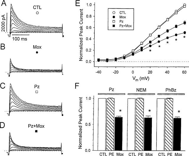
Methoxamine directly blocks outward currents in rat ventricular myocytes. A–D, currents from a representative myocyte during depolarizations from −80 mV to between −20 and +60 mV, in 10 mVsteps at a pulse frequency of 0.2 Hz. Dotted lines denote zero current level. Control (CTL, A) and with 200 μM methoxamine (Mox, B) after 5 min exposure. After 15 min of wash in control solution the cell was exposed to 2 μM prazosin (Pz, C) and then 200 μM methoxamine plus 2 μM prazosin (Pz+Mox, D). (E) mean peak Ito current-voltage relations in control and during exposure to prazosin or methoxamine, or both (n=6). For each cell, Ito was normalized to peak current in control solution measured at +60 mV. Symbols are as marked in panels A–D. * Indicates significant differences between current amplitudes in methoxamine alone compared with both methoxamine and prazosin, P<0.05. (F) block of signal transduction does not prevent the action of methoxamine. Effects of 100 μM phenylephrine (PE) and 200 μM methoxamine (Mox) on Ito amplitude in the presence of 2 μM prazosin (Pz), or after 30 min pretreatment with 50 μM N-ethylmaleimide (NEM) or 1 μM phenoxybenzamine (PhBz), as indicated. Currents were measured at +60 mV, and were normalized to Ito amplitude in the absence of α-agonist, but in the presence of Pz, NEM, or PhBz. Data are means±s.e.mean (n=6). * indicates a significant difference from control, P<0.05.