
Figure 3.
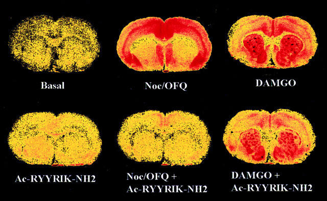
Distributions of basal, 1 μM noc/OFQ- and 5 μM DAMGO-stimulated [35S]-GTPγS binding in absence and presence of 10 μM (noc/OFQ) or 20 μM (DAMGO) hexapeptide Ac-RYYRIK-NH2 in representative sections of rat brain at the level of the caudate putamen. The sections were incubated in Tris buffer/2 mM GDP/100 mM NaCl with 80 pM tracer for 2 h at 25°C.