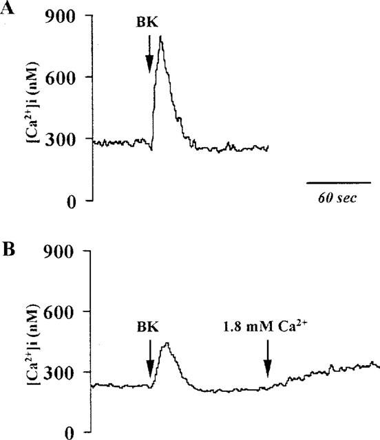
Figure 5.

Effect of extracellular Ca2+ on BK-stimulated changes in [Ca2+]i Trace (A): Cells were stimulated by BK (10 μM) to the buffer containing Ca2+ (1.8 mM). An immediate increase in [Ca2+]i was seen. Trace (B): Cells were incubated in the absence of extracellular Ca2+ and stimulated by BK (10 μM). The response showed a transient increase of [Ca2+]i similar to trace (A) but to a substantially smaller degree. When Ca2+ (1.8 mM) was added, a sustained increase of [Ca2+]i occurred. The traces shown are typical of six separate experiments.
