
Figure 1.
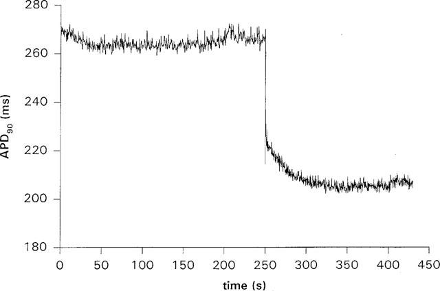
The time course of APD90 shortening during pacing in a guinea-pig ventricular myocyte. The cell was paced at 500 ms CL, and the cycle length was changed to 300 ms after 250 s from the start of recording. On changing the cycle length, APD90 shortened in two stages; a rapid decrease due to electrical restitution followed by a slower exponential decrease lasting about 100 s. The downward spike visible at the start of the slow shortening is due to alternans of APD over two to three beats.