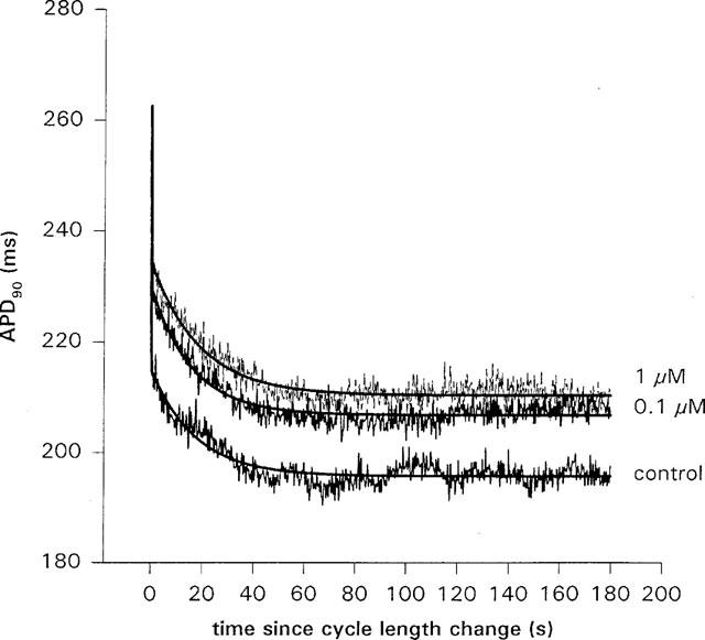
Figure 7.

The effect of dofetilide on the slow shortening of APD90 following a cycle length change from 500 to 300 ms, in an isolated ventricular myocyte. In this cell, the amount of slow shortening, B, obtained from a fit of equation 1 to the data, increased with dose. In control, B=19.5±0.5 ms compared to 23.5±0.5 ms in 0.1 μM, and 24.3±0.5 ms in 1 μM dofetilide. There was no consistent effect on the time constant; τ=17.1±0.7 s in control, compared to 15.9±0.5 s in 0.1 μM and 18.8±0.6 s in 1 μM dofetilide. When the average of four cells was taken, there was no significant change in B or τ with dofetilide dose. Zero on the time axis is the time at which the cycle length was changed.
