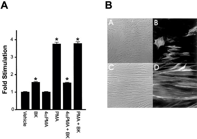
Figure 5.

Effects of PKC activation on PS cells [3H]-thymidine uptake and morphology. (A) Cultured prostate-derived PS cell monolayers were incubated in RPMI 1640, 0.5% (v v−1) FCS for 72 h. Cells were then exposed to vehicle, BK, 4α-PMA (10 nM), PMA (10 nM), 4α-PMA (10 nM) plus BK (10−8 M) or PMA plus BK (10−8 M) for 23 h and pulsed for 1 h with [3H]-thymidine. Incorporation of label into DNA was determined. Data are represented as the mean fold stimulation over vehicle control±s.e.mean for four independent experiments performed in sextuplicate. *P<0.05 compared to vehicle alone. (B) PS cell monolayers in Lab-Tek Chamber Slides were incubated in RPMI 1640, 0.5% (v v−1) FCS for 72 h at which point the various compounds were added and the incubation continued for a further 12 h. Cells were then fixed in methanol and incubated with an anti-smooth muscles α-actin primary antibody and a rhodamine labelled secondary antibody. The labelled cells were observed by phase-contrast (A and C) and immunofluorescence (B and D) optics. Images show cells exposed to 10 nM 4α-PMA (A and B) or 10 nM PMA (C and D).
