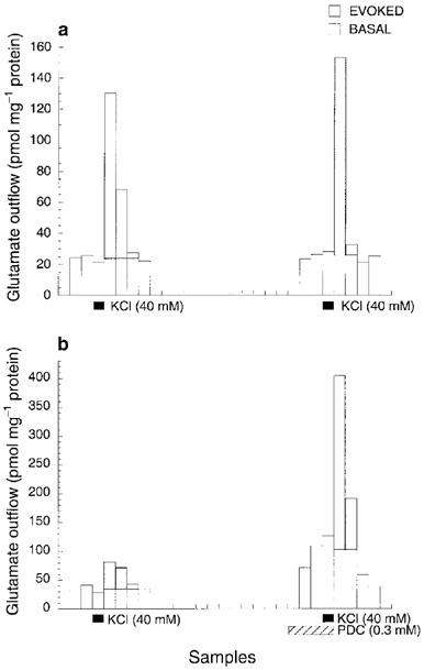
Figure 1.

Representative profiles of glutamate outflow from mouse cerebrocortical slices after two consecutive 1.5-min exposures to 40 mM KCl in the absence (a) and presence (b) of 0.3 mM L-trans-pyrrolidine-2,4-dicarboxylic acid (PDC), a selective glutamate re-uptake inhibitor. All samples were collected in 1.5 min. The time interval between the two KCl exposures was 30 min. The shaded areas represent K+-evoked increases in glutamate outflow above the mean basal level. The integration of the shaded areas is used to calculate the ratio of the second to first K+-evoked glutamate outflow.
