
Figure 1.
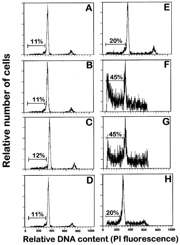
Effects of ET-18-OCH3, BM 41.440 and HPC on the induction of apoptosis in human resting and mitogen-activated T-cells. Resting T-lymphocytes (A, B, C and D) and T-lymphocytes activated with 0.5 μg ml−1 PHA for 4 days and with 0.5 μg ml−1 PHA and 50 U ml−1 IL-2 for 1 additional day (E, F, G and H) were incubated in complete culture medium in the absence (A and E) and in the presence of 10 μM ET-18-OCH3 (B and F), 10 μM BM 41.440 (C and G) and 10 μM HPC (D and H) for 24 h, and then analysed for apoptotic cells by flow cytometry analysis as described in the Methods section. Apoptotic cells distribute at the sub-G1 region of the cell cycle phases. Percentages of apoptotic cells are shown in each histogram. Data shown are representative of four experiments performed.