
Figure 2.
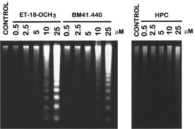
Visualization of DNA fragmentation in activated human T-lymphocytes upon treatment with ET-18-OCH3, BM 41.440 and HPC. Human PBLs were activated for 4 days with 0.5 μg ml−1 PHA and for 1 additional day with 0.5 μg ml−1 PHA and 50 U ml−1 IL-2. Activated T-lymphocytes (2.5×10⁶) were incubated in complete culture medium for 15 h in the absence (control) and in the presence of increasing concentrations of ET-18-OCH3, BM 41.440 and HPC. Then, fragmented DNA was extracted and run onto agarose gels as described in the Methods section. DNA loaded in each lane was from 8×10⁵ cells. Results shown are representative of three independent experiments performed.