
Figure 4.
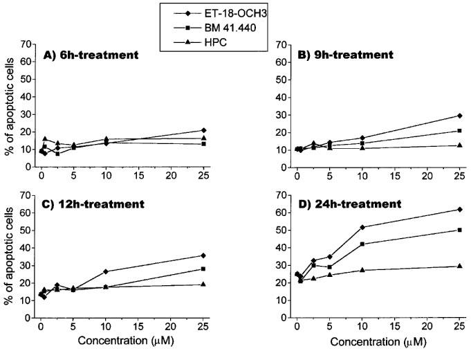
Dose-response and time-course of the effects of ET-18-OCH3, BM 41.440 and HPC on the induction of apoptosis in activated T-lymphocytes. Human PBLs were activated with 0.5 μg ml−1 PHA for 4 days and with 0.5 μg ml−1 PHA and 50 U ml−1 IL-2 for 1 additional day as described in the Methods section. Then, activated T-lymphocytes were incubated with increasing concentrations of ET-18-OCH3 (solid diamonds), BM 41.440 (solid squares) and HPC (solid triangles) for 6 h (A), 9 h (B), 12 h (C) and 24 h (D). Percentages of apoptotic cells were determined by propidium iodide staining of ethanol-fixed cells as described in the Methods section. Control untreated activated T-lymphocytes were also run in parallel. Data are representative of four experiments performed.