
Figure 6.
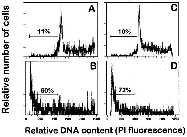
Effect of ET-18-OCH3 on the induction of apoptosis in human leukaemic T cell lines. Human leukaemic T lymphoid Jurkat (A and B) and Peer (C and D) cells were incubated in the absence (A and C) and in the presence of 10 μM ET-18-OCH3 (B and D) for 24 h in complete culture medium, and then analysed for apoptotic cells by flow cytometry as described in the Methods section. Apoptotic cells distribute at the sub-G1 region of the cell cycle phases. Percentages of apoptotic cells are shown in each histogram. Data shown are representative of four experiments performed.