
Figure 7.
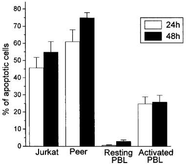
Comparison of the apoptotic action of ET-18-OCH3 on human leukaemic T cell lines versus resting and activated human peripheral blood T-cells. The human leukaemic T lymphoid Jurkat and Peer cell lines, as well as resting and PHA/IL-2-activated PBLs were treated with 10 μM ET-18-OCH3 for 24 h (open histograms) and 48 h (solid histograms) in complete culture medium, and the percentages of apoptotic cells were determined by flow cytometry as described in the Methods section. The percentage of apoptotic cells after ET-18-OCH3 incubation was calculated after subtracting the percentage of spontaneous apoptosis obtained in the corresponding untreated control cells. Data are shown as mean values±s.e.mean from four independent experiments.