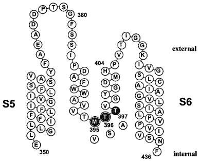
Figure 4.

Scheme of the potassium channel Kv1.3 pore region of one α-subunit. The amino acid sequence of the S5 segment, the pore-loop and the S6 segment is displayed. The position of the mutated internal pore residues are highlighted (gray), with the substitutions shown below (white). The amino acids M395 and T396 are homologous to the part of the internal TEA binding site of Shaker channels and are highlighted by double circles.
