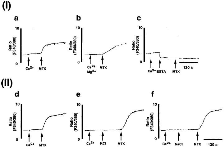
Figure 3.

Maitotoxin-induced extracellular Ca2+-dependent voltage-insensitive Ca2+ influx. (I) Extracellular Ca2+ dependence of maitotoxin-induced Ca2+ influx in C6-BU-1 cells. [Ca2+]i was measured as described in Methods. The cells loaded with fura 2 acetoxy methylester were suspended in the Tyrode solution. (a) Maitotoxin (MTX, 10 ng ml−1) was added 90 s after addition of 1 mM Ca2+ (control for b and c). (b) Maitotoxin (MTX, 10 ng ml−1) was added 90 s after addition of 5 mM Mg2+ and 1 mM Ca2+. (c) Maitotoxin (MTX, 10 ng ml−1) was added after addition of 5 mM EGTA in the presence of 1 mM Ca2+. (II) Maitotoxin-induced Ca2+ influx through voltage-insensitive Ca2+ channel. (d) Maitotoxin (MTX, 10 ng ml−1) was added 90 s after addition of 1 mM Ca2+ (control for e and f). (e) Maitotoxin (MTX, 10 ng ml−1) was added 120 s after addition of high KCl (50 mM) in the presence of 1 mM Ca2+. (f) Maitotoxin (MTX, 10 ng ml−1) was added 120 s after addition of high NaCl (50 mM) in the presence of 1 mM Ca2+.
