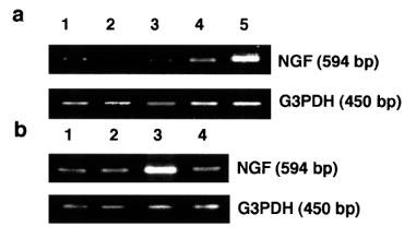
Figure 5.

(a) NGF mRNA expression by maitotoxin. The cells were stimulated by maitotoxin for 3 h under various concentrations of extracellular Ca2+, then total RNA from C6-BU-1 cells was reverse transcribed followed by PCR as described before. Lane 1, control in 1.8 mM CaCl2; 2, control in 1.8 mM CaCl2+3.6 mM EGTA; 3, maitotoxin (10 ng ml−1) in 1.8 mM CaCl2+3.6 mM EGTA; 4, maitotoxin (10 ng ml−1) in 1.8 mM CaCl2+1.7 mM EGTA; 5, maitotoxin (10 ng ml−1) in 1.8 mM CaCl2. (b) NGF mRNA expression by A-23187. The cells were stimulated by A-23187 for 3 h in the presence or absence of 1.8 mM EGTA. Lane 1, control in 1.8 mM CaCl2; 2, control in 1.8 mM CaCl2+1.8 mM EGTA; 3, A-23187 (1 μM) in 1.8 mM CaCl2; 4, A-23187 (1 μM) in 1.8 mM CaCl2+1.8 mM EGTA.
