
Figure 1.
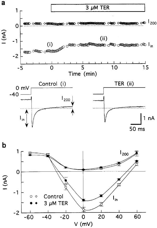
Effects of 3 μM terodiline on membrane currents in guinea-pig ventricular myocytes superfused with Tyrode's solution and dialysed with K+ pipette solution. The myocytes were depolarised for 100 ms from −80 to −40 mV, and pulsed to 0 mV for 200 ms at 0.2 Hz except for determinations of I–V relationships. (a) Data from a representative experiment. Top: time course of changes in peak inward (Iin) and end-of-pulse current (I200) amplitudes at 0 mV, measured with respect to zero current. Bottom: records obtained at the times indicated in the time plot. (b) Average of I–V relationships obtained before (control) and 9–12 min after addition of 3 μM drug; n=five myocytes.