
Figure 5.
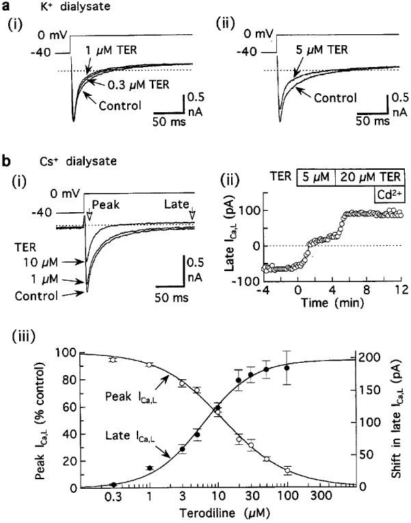
Acceleration of inactivation of ICa,L by terodiline. (a) Records from myocytes superfused and dialysed with K+ solutions, and pulsed at 0.1 Hz. (i) Acceleration in the absence of significant reduction of ICa,L amplitude in a myocyte treated with 0.3 μM terodiline for 5 min and 1 μM terodiline for a further 5 min. (ii) Acceleration in a myocyte treated with 5 μM drug for 3 min. Relatively large outward currents at holding potential −40 mV have been omitted for presentation purposes. (b) Results from experiments in which K+-free solutions were used to minimize K+ currents. (i) Records from a representative myocyte treated for 6 min (1 μM) and then 4 min (10 μM). (ii) Time course of shifts in late ICa,L. (iii) Concentration-response relationships for Ca2+-sensitive peak ICa,L (open circles) and shift in late ICa,L (closed circles). Pulsing rate 0.1 Hz. Number of myocytes: 4–14 at each concentration.