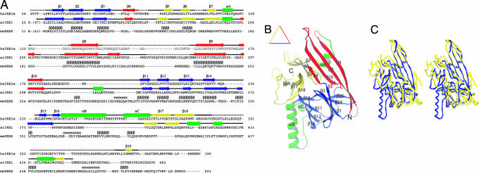
Fig. 1.
The crystal structure of human IRE1α NLD. (A) Sequence and secondary structure alignment of IRE1 and PERK. The NLD sequences of human IRE1α, S. cerevisiae Ire1p, and murine PERK were aligned by using the program T-Coffee (32). Secondary structural elements are indicated above the sequence: α-helices are drawn as rectangles, β-strands as arrows, other elements as solid lines, and structurally unobserved residues as dashed lines. These elements are colored based on their locations in the structure (see Results for details). Predicted α-helices and β-strands for PERK are indicated with Greek letters. (B) A ribbon drawing of the NLD monomer. The secondary structural elements are labeled and colored as in A: α-helices are lettered and drawn as coils, β-strands are numbered and drawn as arrows, and other elements are drawn as tubes. (C) A stereo diagram showing Cα trace superimposition of human IRE1α NLD (blue) and S. cerevisiae Ire1p NLD (yellow) structures. The programs Ribbons (33) and Grasp (34) were used to produce B and C, respectively.

