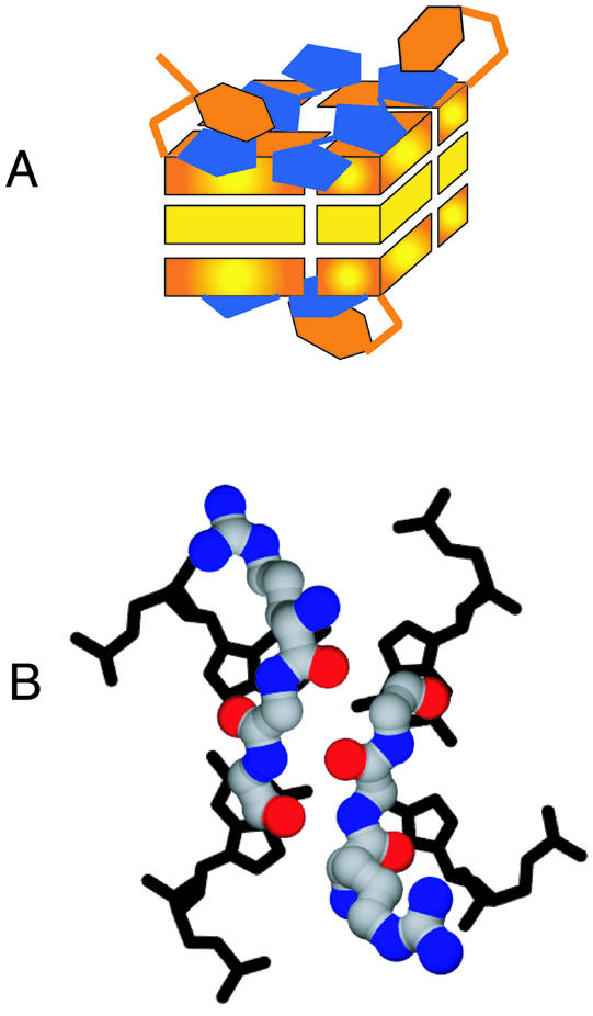
Figure 6.

Models for interaction of distamycin and RGG repeats with G4 DNA. (A) A binding mode which allows two distamycin molecules to dock onto each terminal G-plane. Distamycin bound as shown would inhibit protein binding by occluding the terminal G-planes. Unfortunately, owing to the symmetry of the DNA and because the ligand is in fast exchange between multiple conformations with relatively few NOEs, the data are insufficient to derive a single structural model for binding. This is one model consistent with the NMR data. (B) Speculative model for the interaction of two protein RGG units with G4 DNA. The two RGGs appear in a short β-sheet conformation with the arginines positioned to form salt bridges with DNA phosphate groups.
