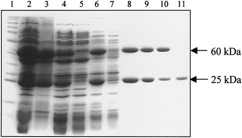
Figure 1.
Purification of M.AhdI. SDS gel electrophoresis showing the purification of the methylase complex. Lane 1, molecular weight marker; lane 2, mixed cell lysates; lane 3, insoluble fraction; lane 4, soluble fraction; lane 5, supernatant after ammonium sulphate precipitation. Lanes 6–8, heparin column; lane 6, sample loaded; lane 7, run-through; lane 8, pooled fractions. Lanes 9–11, gel filtration; lane 9, sample loaded; lane 10, methylase peak; lane 11, S subunit peak.

