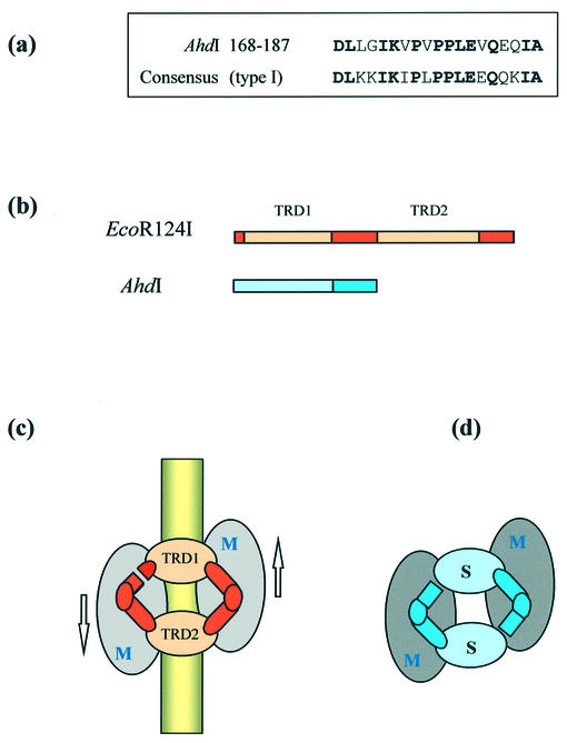
Figure 7.
(a) Sequence alignment of residues 168–187 of the S subunit of AhdI and the consensus sequence found at the start of the central conserved region of all type I S subunits. (b) Comparison of the sequences of the S subunits of a type I MTase (EcoR124I) and AhdI. Conserved sequences (darker colours) are repeated in type I S subunits and include a limited region of sequence homology shared by all type I MTases. TRDs (lighter colours) responsible for recognition of DNA half-sites are unrelated in sequence. (c) Circular model for the subunit/domain structure of type I enzymes of the form M2S1 [adapted from Kneale (6)], showing the approximate two-fold symmetrical disposition of subunits and the orientation of the enzyme on the DNA. (d) Proposed symmetrical model (M2S2) for M.AhdI.

