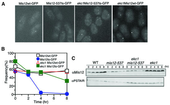
Fig. 4. Restoration of Mis12 mutant protein localization in the ekc1-163 background. (A) The dot-like localization of Mis12 mutant protein was restored in the double mutant mis12 ekc1. Images represent Mis12–GFP and Mis12-537–GFP expressed in the wild-type and ekc1 background cells (36°C, 8 h). (B) The frequencies of the centromeric dot-like appearance of Mis12ts–GFP were restored in ekc1 mutant cells. Mis12ts–GFP signals in the wild-type background became diffused after 4 h. (C) Schizosaccharomyces pombe cell extracts of wild-type and mutant cells cultured at 36°C were prepared at 2 h intervals. Immunoblotting of extracts was performed using anti-Mis12 and control anti-PSTAIR antibodies.

An official website of the United States government
Here's how you know
Official websites use .gov
A
.gov website belongs to an official
government organization in the United States.
Secure .gov websites use HTTPS
A lock (
) or https:// means you've safely
connected to the .gov website. Share sensitive
information only on official, secure websites.
