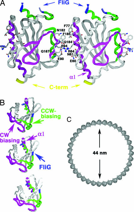
Fig. 5.
Assembly of FliM in the C-ring. (A) FliM self-association model based on cross-linking data, functional analyses, and intersubunit spacing within the C-ring (see Figs. 3 and 4). FliM self-associates through interactions mediated by largely hydrophilic side chains of α1, β1′, and the α2′–α2 region on the opposing subunit. The GGXG motif implicated in binding FliG is partially disordered (blue). The C terminus of the molecule projects from the bottom to interact with FliN. (B) Top view of three FliM subunits in the C-ring. Switching may involve rotation of the subunits to place the CCW-biasing patch (green) within the subunit interface. (C) An assembly of 35 subunits would generate a C-ring of diameter 44 nm.

