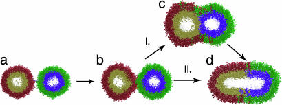
Fig. 2.
Branching reaction pathway for vesicle fusion. Pathway I shows the canonical progression from an unfused starting state (a) through a stalk-like early intermediate (b) and a hemifused late intermediate (c) to the fully fused state (d). Pathway II shows the additional reaction pathway observed in our simulations: rapid fusion from the stalk-like intermediate to the fully fused state. All renderings are of snapshots from observed reaction trajectories; lipids are colored to distinguish the outer (red and green) and inner (gold and blue) leaflets of each vesicle. Explicit water is present in all simulations but not rendered.

