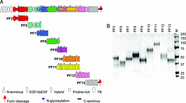
Fig. 1.
Schematic diagram of the domain structure of fibrillin-1 and the protein fragments used in this study. (A) The domain structure of fibrillin-1 is shown colored by protein fragment. All expressed fibrillin-1 fragments are shown below their position in the fibrillin-1 molecule. A striped representation on the fibrillin-1 molecule indicates overlapping fragments. A key indicating the different domains, N-linked glycosylation sites, and C-terminal furin cleavage site is shown. The protein fragments are shown in these corresponding colors throughout the figures. (B) SDS/PAGE showing fibrillin-1 fragments, PF2, -5, -7, -8, -9, -10, -11, -12, and -13, before HPLC. M shows standard molecular weight markers.

