Fig. 3.
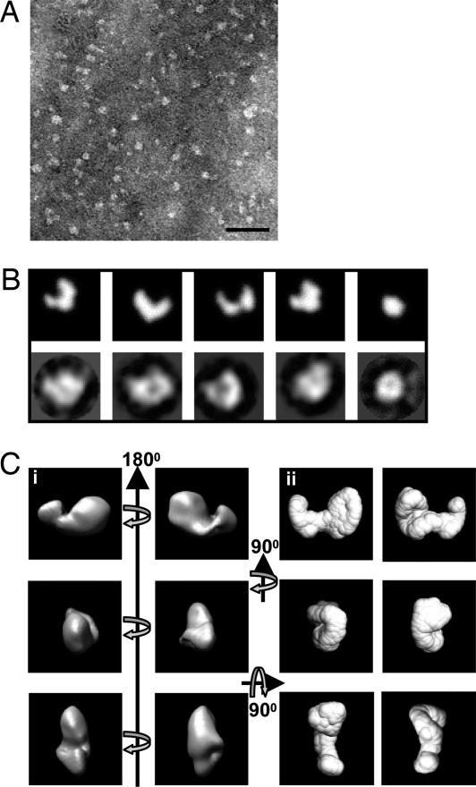
Comparison of the structures of PF11 determined by single-particle TEM and solution SAXS. (A) Negatively stained TEM image of fibrillin-1 fragment PF11 recorded on a Tecnai12 Twin at 120 keV. (Scale bar: 50 nm.) (B) (Upper) 2D projected images from the ab initio SAXS structure of PF11 were calculated by using IMAGIC. (Lower) These were compared with representative class averages from single-particle image processing of PF11. There are striking similarities in the size and shape of PF11 when compared by the two techniques. The class averages were generated without a reference and are completely independent of the SAXS structure. (Ci) The 3D reconstruction of PF11 was calculated by using angular reconstitution and is shown here in orthogonal views as a volume-rendered representation. (Cii) For comparison, the ab initio SAXS structure was drawn as a surface representation and shown in the same orientations. Again, there are very clear similarities between the single-particle EM and SAXS structures of PF11. For B and C, the boxes are 14 ×14-nm square.

