Fig. 6.
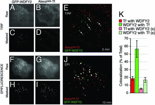
Trafficking of Tf through WDFY2-containing endosomes. (A–J) HeLa cells stably expressing GFP-WDFY2 were incubated with fluorescent transferrin (Alexa568-Tf) and imaged by TIRF (A–E) and epifluorescence (F–J). TIRF images were obtained after 5 min, and epifluorescence images of the same cell were obtained after 10 min of continuous exposure to Alexa568-Tf. Raw (A, B, F, and G) and masked (C, D, H, and I) images of GFP-WDFY2 (A, C, F, and H) and Alexa568-Tf (B, D, G, and I) are shown. Colocalization between GFP-WDFY2 (green) and Alexa568-Tf (red) is rendered in white in overlaps of the masked images (E and J). Arrows point to colocalized voxels in the TIRF images. (K) The colocalization between Tf and WDFY2 in TIRF images from cells incubated for 10 min in the continuous presence of Tf was quantified. Rectangular areas enriched in both signals were analyzed, and colocalization was expressed as the percent of the total fluorescence from Tf colocalizing with WDFY2 and vice versa. Spurious colocalization was obtained by flipping one of the images along the x axis. Bars and vertical lines represent the mean and standard error of the real (red and green) or spurious (light red and light green) colocalization measured in 10 regions from 10 independent cells. (Magnifications: A–D, ×1,000; F–G, ×2,000.)

